Four Graduate Students Win Briscoe Library’s Image of Research Photography Competition
Four graduate students have won prizes in the Briscoe Library’s Image of Research competition. The awards are sponsored by the Office of the Vice President for Research and Academic Faculty, and Student Affairs.
According to the website, “The Image of Research competition is an opportunity for UT Health San Antonio students from all six schools to capture, share, and present the essence of their research in a single visual image. This competition and its accompanying exhibition showcase students’ creative visual conceptualization of their research.”
1st Place: Jaclyn Merlo
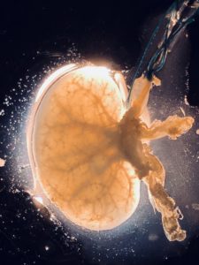
Rodent Kidney Extracellular Scaffold
The image presented is of a de-cellularized rodent kidney displaying the collagen matrix of the renal vasculature, tubules, and glomeruli. Rapid de-cellularization is accomplished by perfusing a surfactant solution through the renal artery, under exposure to an electric field within a bioreactor. The novel bioreactor, developed at UT Health San Antonio, removes resident cells ten times faster than by traditional de-cellularization technology while preserving elements of the matrix that are critical to directing stem cell differentiation.
High-quality extracellular scaffolds are indispensable for research in regenerative medicine, gene transfer, cancer, and tissue transplantation. The extracellular scaffolds of specific animal tissues can provide templates for the differentiation of human stem cells for the study of diseases in more relevant models, thus facilitating translation to human medicine. Further, the technology is scalable and can prepare large animal and human tissue extracellular scaffolds.
About Jaclyn
 Jaclyn is a native of San Antonio and a first-generation college student. She graduated from Texas Tech University in 2015 with a Bachelor’s degree in Chemistry. Even though her background is in chemistry, she fell in love with the biomedical sciences in her senior year as an undergrad. She is currently in the pursuit of becoming a physician scientist and she is in the Integrated Biomedical Sciences program in the Molecular Immunology and Microbiology discipline at UT Health San Antonio. She is currently a member of Dr. Leon Bunegin’s lab, studying regenerative medicine and limb preservation.
Why did you choose this image?
“I chose the image because, at first glance, its aura is alluring and represents the possibilities of the rodent kidney as a template for human stem cells. A single light was shown through in complete darkness, and created the overwhelming detail of the scaffold’s natural beauty on a microscale.”
3rd Place: Camila Pereira
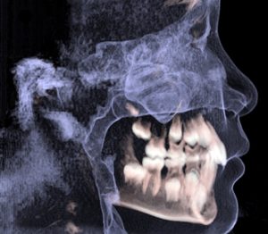
Airway Space Tour – A 3D Ride
The airway should be free of obstacles such that air can follow its course from the nasal cavity into the lungs. Our research investigates the airway space imbalance that affects children who breath through their mouth while sleeping. Dental 3D radiograph should be used as opportunistic screening tool for sleep-related breathing disorders such as snoring and sleep apnea. These disorders could be caused by hypertrophied tonsils and nasal obstruction between others. Due to the lack of good sleep, children could have low grades at school, difficulty to concentrate, and disturbed cognitive abilities. Other signs such as delayed growth, tiredness, irritability, or lack of energy even to play are related. Ultimately, 3 dimensions of life are affected: craniofacial growth, intellectual development and quality of life. When the dysfunction is detected early enough, the consequences can be reduced or even eliminated. We hope the translation of our research project will increase awareness and raise the attention of the dental professionals’ and the general public to this matter. The sleep disordered breathing is a public health issue and surveillance is essential. Let’s take this ride!
About Camila
 Camila Pacheco Pereira was born and raised in Brazil. She has lived in Canada for more than 10 years. She is in the Master of Science in Dental Science program specifically in the Oral Maxillofacial Radiology track.
Why did you choose this image?
The image reflects a 3D reconstruction of a cone-beam computed tomography – a dental radiograph that we see on a daily basis. The mixed dentition and the airflow can be easily identified by the general public. The image represents well the reason behind why we should be vigilant to the craniofacial growth of our patients on a bigger scope. Signs of asymmetry, imbalance, or dysfunction should not be neglected.
IPE Award
Sarah Khoury, Cell Systems & Anatomy
Daryl Gaspar, Pharmacy
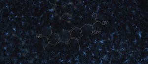
Treatment in the Stars
Astrocytes carry great potential for stroke treatment and research conducted in the past has generally ignored their ability to heal neurons. Research suggests that use of fatty acid oxidation by astrocytes may be useful for healing, and protecting tissues that have been affected by stroke. Triiodothyronine (T3), a thyroid hormone, stimulates fatty acid oxidation, stimulating the production of ATP in astrocytes. In mice treated with T3 stroke lesion volumes are smaller than those without treatment. In this image the brighter activated astrocytes indicate a stressed brain, one that has experienced an injury. T3, the constellation found in the middle of the image may one day be used for stroke treatment.
