Final Words: John Walker Explains How to Balance Life in Graduate School
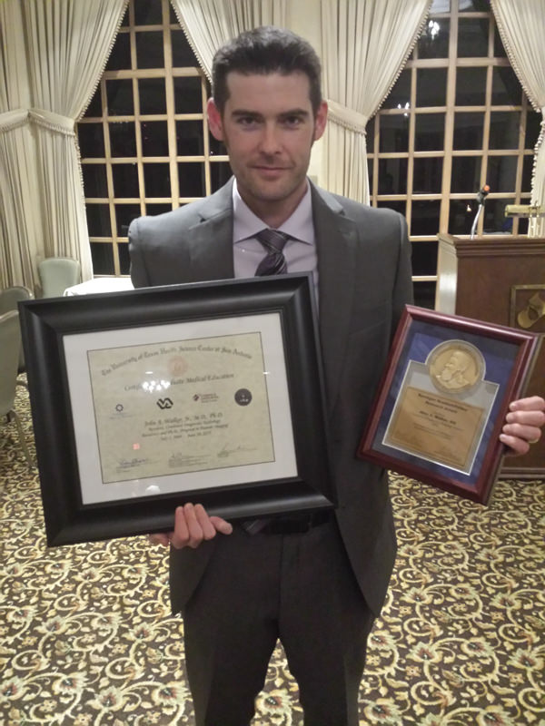
Congratulations John Walker in the Radiological Sciences-Human Imaging program for successfully defending your dissertation on “Computed Tomography Perfusion for Assessing Vascularization in Small Animal en bloc Tissue Engineering Models.”
Please tell me about yourself, why did you pick UT Health Science
Center, and your program.
 I have been part of the UT Health Science Center at San Antonio since 2003, when I started medical school.
Following graduation, I expanded my horizon through a two-year commitment to research in a partnership between the Department of Surgery here at the HSC and the U.S. Army Institute for Surgical Research.
At that time my interests were application of tissue substitutes,
but that led to an interest in imaging in hopes of better understanding
substitutes integration with native tissue, in particular vascular integration
that is vital for cell survival.
Why are you passionate about your research topic? How did you first become interested in it?
My passion for helping advance the field of tissue regeneration lies in the fact that I cherish quality of life even beyond that of quantity.
When you see individuals with deficits related to lack of tissue, be it a failing organ or loss of limb, such that their capacity to live life to the fullest is hindered, then the reward of giving someone back that ability is priceless.
I do however get an added bonus by placing my efforts into imaging, in that I get to play with really advanced
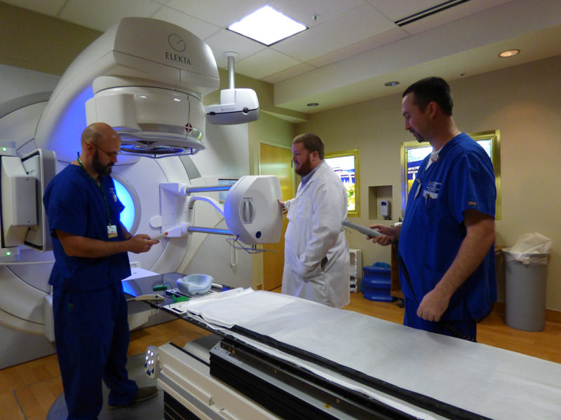
technology. I always kind of knew I was a closet techy, but once I was introduced to imaging during my initial research experience, I could not get enough.
So, I altered career paths and entered the field of radiology, where I will now get to spend the rest of my medicine career being a giant kid with unbelievable fun toys, all aimed at helping others gain a higher quality life.
Please provide a few sentences summarizing your dissertation. What was the experience like for you?

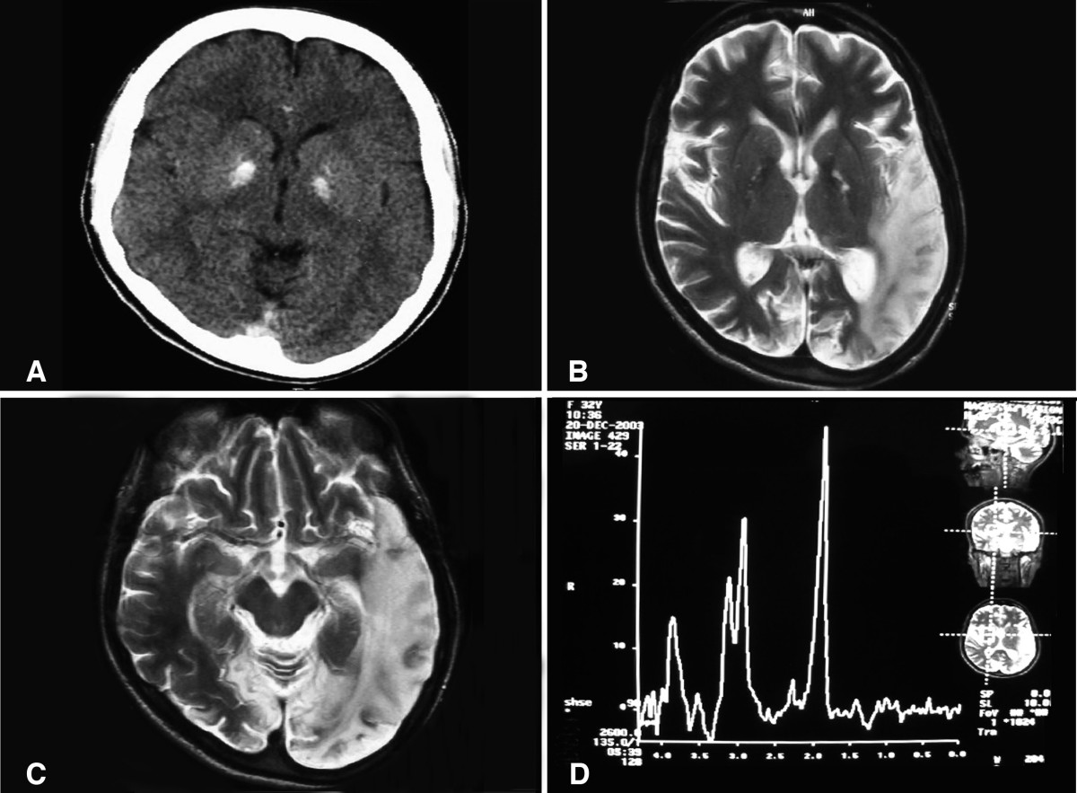
My dissertation adapts current dynamic enhanced computed tomography techniques that measure perfusion parameters among clinical stroke, and experimental heart and tumor models into small animal implants.
Two important considerations to this work included, small size and fast physiology. The argument for my work was that our currently
advanced multislice clinical CT scanners could provide appropriate spatial resolutions among subcentimeter implants at temporal resolutions necessary to calculate meaningful perfusion parameters.
The first study focused on defining blood flow and blood volumes among normal rat anterior tibialis muscles at rest and with variable degrees of stimulation provided to alter muscle perfusion. We were able to show predictable blood flow and volumes based on degree of stimulation by our imaging techniques.
Our second study expanded on imaging the rat anterior tibialis muscle by implanting a 6 mm tissue engineered construct composed of collagen and microvascular fragments into the rat anterior tibialis muscle. The experimental aim was designed to challenge the spatial resolution while answering whether the known improvement in histological vessel density equated to in vivo perfusion, and to what degree if so.
Furthermore, the experiment used stimulation to alter perfusion dynamics to improve spatial resolution by providing differing blood flow contrast to background muscle, but to further understand the interplay of implant perfusion with native host muscle perfusion. A third, paralleling experiment, challenged the technique among various degrees of mineralizing subcentimeter bone implants.
This was performed to better understand at what degree mineralizing implants may no longer show reliable perfusion measurements, an important concept given other modalities are poorly suited to image bone, and also to characterize the interplay between perfusion changes and mineralization.
Overall, the experiments showed promising results to conclude that the imaging resolutions are sufficient to ascertain extent and distribution of blood flow within implanted tissue substitutes among our small animal models, to include mineralizing implants less dense than native bone.
What was your best memory during graduate school or what did you learn?
 I really think graduate school provided me a lesson in balancing life.
As a dual resident and graduate student, I had to begin planning far into the future so that I could design protocols that would allow
me to maximize data collection while tending to clinical radiology.
This turns out to be a more challenging task than the surface reflects, because to effectively design protocols to fit schedules, the
totality of the experiments has to be envisioned so that pitfalls can be troubleshot prior to arising.
In the beginning, I was weak in this area and failure to streamline delayed any seemingly usable data for the first year and half after I began running experiments.
But as I began to learn the skill, I was able to make up for lost time as protocols began to parallel one another and seemingly happen on less personal time.
Life and career skills such as these are invaluable. I must also add that graduate school afforded me time to continue working with many colleagues that continue to enrich my life and I hope to continue working with for years to come. Great colleagues are priceless.
What’s next?
Immediately in my future is fellowship training as a fellow in Vascular and Interventional Radiology, here at the HSC (who could ever leave this place?).
Regarding research, I hope to garnish an academic position at the conclusion of my fellowship, and hope to continue working on improvements among tissue substitutes along with ways to improve outcome assessments through imaging.
I foresee a day when the interventionist can use minimally invasive techniques to either strategically place substitutes and/or improve survivability of implanted substitutes, and I hope that I get to be part of that chapter in history.
Any advice for your fellow graduate students?
While knowledge is a tool that is necessary to move forward, curiosity, and perseverance will provide you with the knowledge needed to expand on what is already.
So never stop being curious, never stop trying, for all problems beg to be solved, but with each solution will come new questions.
I wish you good luck in this never-ending field we call research!
