A 3D Model To Help Students Understand Anatomy Better
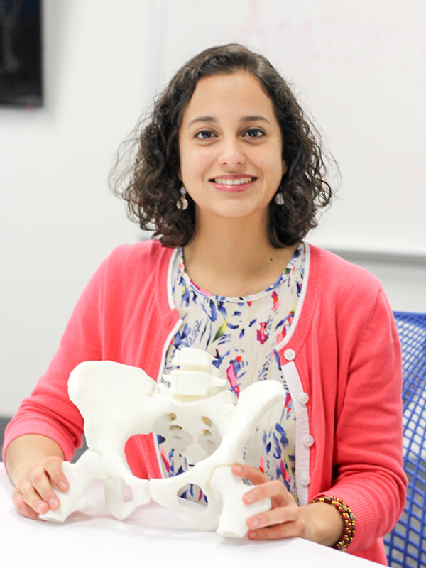 Master of Science in Cell Systems and Anatomy student Laura Solis was recently featured on the Health Science Center’s library for her innovative thesis project.
Laura decided to use the Briscoe Library’s 3D printing service to help students understand anatomy especially of the pelvis and perineum regions.
“3D printing has existed for 30 years and its use in education has provided access to novel and interactive teaching tools for students” she said. “In the health sciences, the anatomy of the pelvis and perineum is very difficult to understand because of the multilayered arrangement of structures within a narrow bony space. Students find it very challenging when dissecting these regions in the gross anatomy lab and the two-dimensional pictures in the atlases and books tend to simplify the complexity of the anatomy.”
The 3D printed structure, which is the bony base of Solis’ model, is the solid replica of a digital file made by other scientists. This CAT file is available online to the public and can be downloaded for free.
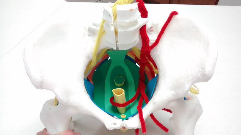
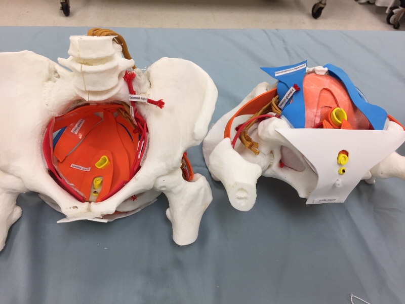
“By printing the bony pelvis in 3D, I was able to build the inside of the pelvis and the perineum layer by layer with the ligaments, muscles and vessels made with craft materials including foamy, and para-cord with wire. Most importantly, this model showed important spatial relationships that student have difficulty grasping during their first exposure to the anatomy.”
Solis explained that each full model took around 58 hours to make. Eleven built models were used during a medical student’s gross anatomy lesson on the pelvis and perineum.
“Students divided in teams participated in an interactive lesson on the pelvis and perineum. Each team had a built-in pelvis, which they were able to disassemble since Velcro was used to attach the structures to the bony pelvis. The students had a handout with an activity that reviewed the structures and asked questions about their relationships” she said. “Although this model could directly benefit future OB/GYN doctors, it is an example of a teaching tool that could adapt to other anatomical regions and for other health professional students.”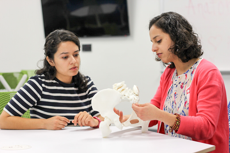
As part of the project, she also helped on the creation of a clinical case of the pelvis and perineum that used BodyViz software to show abnormal and normal anatomy in a clinical context.
“The BodyViz software showed students the radiographical view of the anatomy” she said. “As physicians, students will look at MRIs, CT scans, and X rays so the clinical case introduced a clinical application. I think this has been a good project and hopefully will make an impact on student learning by improving their understanding of important spatial relationships.”
Laura plans to use the 3D model in future conferences and feels grateful to her research mentor, Dr. Omid Rahimi and the Cell Systems and Anatomy faculty.
“I greatly appreciated the opportunity that Dr. Rahimi granted to present my model to medical students, and I have truly valued the support I have received from him and the members of my committee. Although there is still work to be done, I have enjoyed the experience and feel confident that the project will be soon completed.”
After graduation, Solis plans to apply to medical school.
“The human aspect of medicine calls to me. My interest is to work with underserved populations in their health education and disease prevention. As I have shadowed physicians, I’ve seen how much their patients trust them and it’s empowering to see how grateful patients are with their work.”
Close Ups of The Model
 This article was written by Charlotte Anthony, marketing specialist at the Graduate School of Biomedical Sciences at UT Health San Antonio. This article is part of the “Meet The Researcher” series which showcases researchers at the Graduate School of Biomedical Sciences at University of Texas Health Science Center San Antonio.
